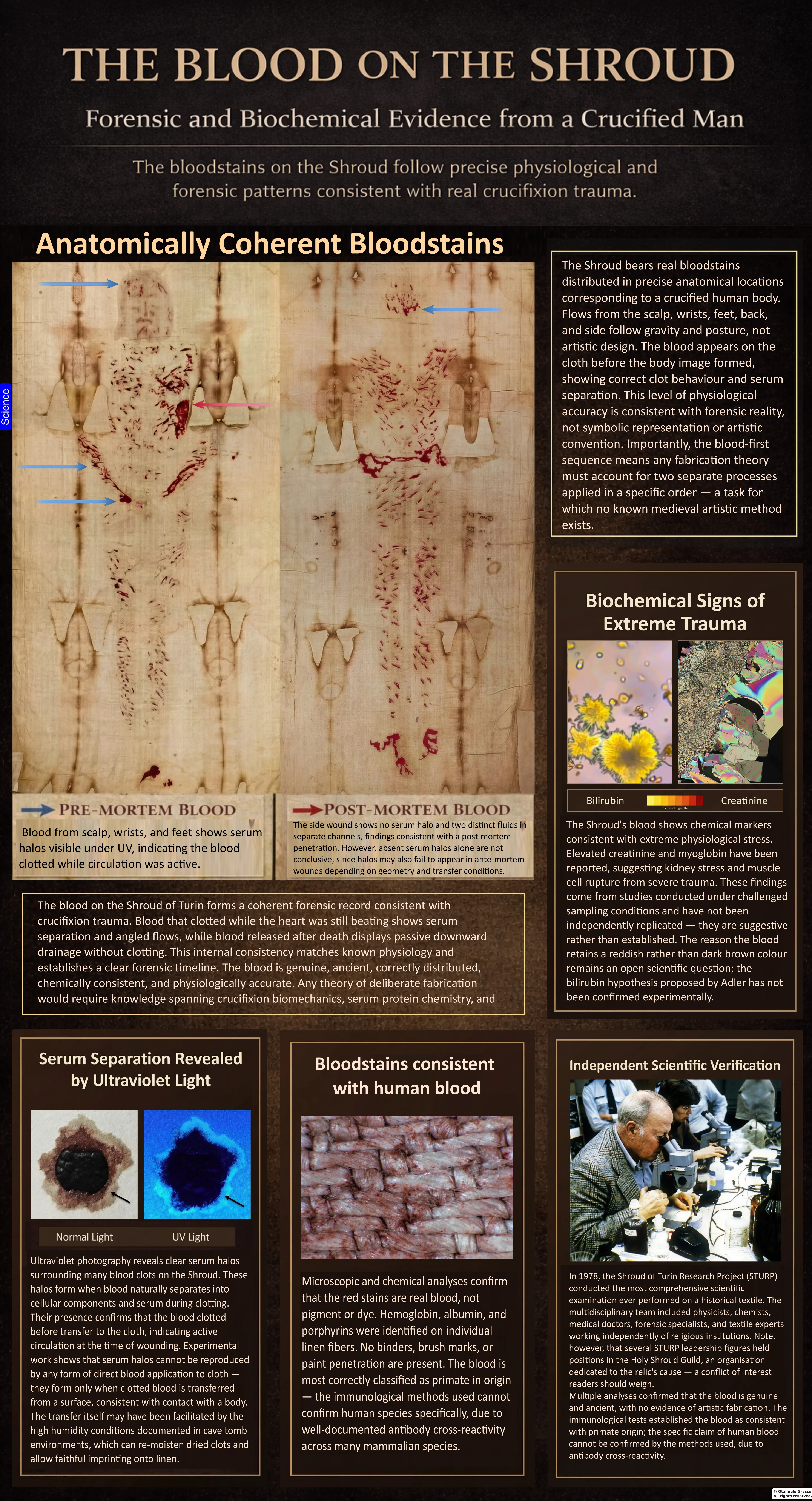
A CLOSER LOOK · BLOOD MARKS
The Blood of the Shroud
The reddish marks on the Shroud have been called blood for centuries. Modern investigators asked a narrower set of questions: what can chemical tests identify in tiny samples; how do the marks behave under visible and ultraviolet light; and can experiments explain how blood and clear fluid might have reached linen? Those questions have produced important evidence and real disagreement. They do not, by themselves, identify the person, date the cloth, or reconstruct one certain sequence of events.
This chapter follows that evidence from the first laboratory tests through the 1978 examination and more recent transfer experiments. It also revisits stronger assertions in the author’s earlier book in light of the later critical review supplied for this project. Reported laboratory observations, modern experimental analogues, forensic interpretations, and historical or theological conclusions are kept separate throughout.
1. The questions the stains raise
The Shroud bears reddish-brown marks near the scalp, face, wrists or hands, feet, side, back, and legs. Their locations have long been interpreted in relation to wounds from scourging and crucifixion. The visible pattern is a starting observation, not a conclusion about what material made it or how it was deposited.
At least five questions must be answered separately. First, do the marks contain blood-related material? Second, can its species, blood group, or biological source be identified? Third, what do stain shape, direction, and ultraviolet behavior reveal about fluid separation and transfer? Fourth, can those observations distinguish bleeding during life from drainage after death? Fifth, do any of those answers identify the cloth or the person represented? Evidence for one question does not automatically answer the next. A blood-like mark may contain genuine blood yet have been deliberately applied; a serum-like margin may be consistent with clot separation without proving direct contact with a corpse; and a plausible wound reconstruction does not establish an identity.
This distinction matters because some statements repeated in popular and devotional accounts—“human blood,” “type AB,” “male,” “arterial,” “postmortem,” “serum halo,” or “proof of crucifixion”—sound like laboratory labels. In fact, each involves methods, assumptions, and limitations. The appropriate standard is to state what was observed, what a test can detect, and what alternative explanations remain.
2. From the 1970s tests to the present
In 1973, a Turin commission authorized examination of a few threads from marked areas. Giorgio Frache, Eugenia Mari Rizzati, and Emilio Mari reported negative results from hematological tests, including benzidine-based procedures and microspectroscopic examination. They did not claim that a negative result absolutely excluded ancient blood: the material was minute and degraded, and a negative test on a particular thread is not a test of every stain on the cloth. This early result belongs in the history because it shows that the composition was not settled by visual inspection alone.1
In 1978, the Shroud of Turin Research Project (STURP) conducted a five-day multidisciplinary examination. The team photographed the cloth under visible and ultraviolet light, collected adhesive-tape samples, and used spectroscopy, microscopy, and other instruments. Biophysicist John Heller and chemist Alan Adler then analyzed tape samples from areas interpreted as bloodstains. They published a 1980 report in Applied Optics and a longer chemical study in 1981. The studies reported evidence they interpreted as blood-related, including heme-derived spectra and protein reactions. These reports became central to the affirmative case, but the samples were small and the findings have not all been independently replicated using current forensic protocols on authenticated stain material.2, 3, 4
STURP’s broad examination also included ultraviolet fluorescence photography by Vernon Miller and Samuel Pellicori. Their images recorded pale fluorescent margins around some reddish stains. These photographs are direct historical data that can be inspected; calling every margin “serum” is a further interpretation that requires comparison with controlled samples. The original ultraviolet record has not been comprehensively re-imaged with modern calibrated equipment.5, 6
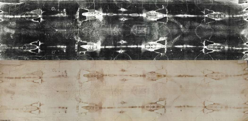
Walter McCrone, a microscopist who examined tape samples, argued that particles he identified as iron oxide and vermilion supported an artistic-pigment explanation. Heller and Adler challenged that interpretation, arguing that the observed chemistry and particle distribution were not enough to explain the marks as paint. The disagreement is not resolved by saying one investigator “saw iron” and another “found blood”: iron-bearing particles can have multiple origins, and blood residues can coexist with other material on a handled, repaired textile. The central issue is whether each method and sample can explain the particular marks being discussed.7, 8, 9
A 2016 review by hematologists Gian Franco Di Minno and colleagues cautioned against treating older blood identification tests as conclusive by modern standards. They emphasized that aged material, assay specificity, contamination, and cross-reactions complicate interpretation. Their review does not show that the stains are pigments; it does show why the strongest claims should not outrun the validation of the tests.10
3. What can be seen on the cloth?
At ordinary scale, some marks appear as compact blotches, some as narrow downward tracks, and others as broader transfers or irregular patches. Their distribution is compatible with a body that suffered injury, but geometry alone does not name the fluid. Lighting, camera response, image processing, weave texture, stains, repairs, and the 1532 fire and its extinguishing water can all affect apparent color and edge shape. A researcher should distinguish the original observation from the label placed on it.
The head region is often described as showing numerous punctures and rivulets; the back and legs as repeated scourge marks; the wrists and feet as flows associated with nail wounds; and the side as a larger wound with blood and a pale margin. These are anatomical interpretations, not direct measurements of instrument, vessel type, arterial pressure, or exact posture. Terms such as “arterial” and “venous” require diagnostic features and validated comparison, not color alone. The figure used on the former page carried categorical labels that the available stain photograph cannot establish, so this expanded article relies on unannotated source imagery and clearly qualified captions instead.
It is also important not to assume that all marks have one history. A cloth can bear original stains, later transfers, handling residues, conservation effects, and fire or water damage. A genuinely mixed set of materials or deposition episodes is possible. The forensic task is to map and analyze marks individually, then ask whether a coherent sequence explains them better than alternatives.
4. The chemistry debate: what has been reported?
Heller and Adler used microspectrophotometry and chemical reactions on adhesive-tape material from selected stained areas. They reported spectral behavior consistent with heme compounds and tests interpreted as supporting hemoglobin-derived material. In the 1981 paper they also described protein reactions, including albumin, and other chemical observations. STURP publications combined these with observations of stain distribution and the absence, in the tested fibers, of a pigment layer they considered sufficient to explain the body image. Their conclusion that blood components were present is a significant published result, but “the samples tested showed blood-related signatures” is more precise than “every red mark has been definitively identified as human blood.”2, 3, 4
Different tests have different levels of specificity. A color reaction may indicate a chemical class but can have interfering substances. A spectrum consistent with heme is not automatically a species identification. A protein assay is only as specific as its antibody and controls. Iron detection alone does not prove hemoglobin because iron occurs in pigments and environmental particles. A persuasive identification should show the method, controls, sample origin, sensitivity, possible cross-reactions, and independent replication.
McCrone’s microscopy identified iron-rich and red pigment particles in some tape material and led him to argue for artistic application. Fanti and Zagotto later proposed that pigments might reinforce blood staining. These findings are relevant to the debate but do not necessarily explain every mark or establish that the stains are wholly artificial. Conversely, a blood-related signal in a few samples does not establish when it was deposited or whether it came directly from the depicted body. A fair assessment must allow mixed composition and evaluate evidence at the location actually sampled.8, 9, 11
There is a further limitation: the 1978 work used tape lifts, not a modern, geographically distributed sampling plan. Tape can collect surface material and fibres, but does not provide a complete cross-sectional record of every layer. Fire, water, conservation, devotional contact, and historical handling complicate preservation. The results are technically interesting and historically important; they are not equivalent to a modern, independently replicated forensic identification from a chain-of-custody sample.
The author’s earlier biochemical synthesis also discusses bilirubin, urea, and creatinine as possible traces of severe trauma, dehydration, or blood breakdown. These are hypotheses about tiny, old, heterogeneous samples; they should not be presented as a medical diagnosis of the depicted individual. A 2022 study by Kearse examined ultraviolet fluorescence associated with elevated bilirubin in modern dried serum, while Di Lascio and colleagues tested hypotheses about the color of archaeological bloodstains. Neither study independently confirms the claimed concentration or clinical meaning of a particular Shroud stain. The author’s later review grades ferritin/creatinine and similar trauma-marker claims as insufficiently validated for strong conclusions. Those measurements remain interesting leads requiring independent confirmation and contamination controls.20, 35, 36
5. Species, sex, ABO group, and DNA
Species. Earlier reports described reactions interpreted as human or primate blood. Kelly Kearse’s review of species testing explains that polyclonal “anti-human” antisera can cross-react with proteins from other species and that degradation further complicates ancient samples. In modern forensic practice, a robust species identification needs validated, specific assays and appropriate controls. The available legacy results do not establish human origin to contemporary forensic standards; the careful conclusion is that the species remains unconfirmed.12, 13
ABO group. The often-repeated AB designation traces principally to older serological work by Pierluigi Baima Bollone and collaborators. Kearse’s 2023 review details why ABO typing of ancient artifacts is unreliable: antigens can degrade, environmental substances can react with test reagents, and results from aged material cannot be interpreted like a fresh clinical sample. The historical report should be mentioned as a claim in the literature, not stated as a confirmed blood type.14, 15
Sex and DNA. No reliable test has established the sex or genetic identity of the person who contributed the stain material. The Shroud has been handled, displayed, repaired, sampled, and exposed to many environments. DNA recovered from an ancient surface can represent handlers, conservators, visitors, or environmental transfer. Even DNA on a fibre near a stain is not automatically DNA from the blood or from the man represented. Kearse’s review of aged-blood DNA methods stresses provenance, contamination controls, validated extraction, replication, and careful interpretation. Claims that the blood is “male,” “Semitic,” or genetically matched to another relic exceed what is established.16
These limits do not show the blood is not human, not AB, or not from a male. They mean that the evidence currently available does not justify presenting those identifications as facts.
6. The proposed sequence between blood and body image
Heller and Adler argued that blood and body-image coloration behave differently on the linen, and that image coloration is absent or reduced beneath some blood material. They interpreted the stratigraphic relationship as evidence that blood was present before the body image formed. The author’s earlier book calls this a “first law” of Shroud forensics and treats it as a major obstacle to a medieval painting scenario. This is an important hypothesis, but its evidential reach depends on how the particular fibres were sampled, how their surface layers were examined, and whether the relation is consistent across the cloth.3, 4, 17
A microscopy or tape sample can show a local relationship; it cannot establish a complete sequence for every stain without mapped, independently replicated cross-sections. It also cannot by itself exclude a maker who applied genuine blood before adding an image, or who used multiple materials and stages. The exact tests and the local sequence should be described, rather than turning a reported sample observation into a universal rule or an authentication result.
The distinction is important: a layer order can constrain a proposed process, but it does not date the layer or identify who made it. The most useful next step would be high-resolution, non-destructive mapping of blood-associated marks and image fibers, paired with controlled laboratory analogues and clearly documented limitations.
7. Ultraviolet margins: observation, serum, and alternatives
Under ultraviolet illumination, some stains show a paler fluorescent margin beyond a darker center. Miller and Pellicori recorded such features in 1978. “Fluorescent margin” describes what the photograph shows. “Serum halo” proposes what that margin is. In clot retraction, platelets and fibrin contract and expel liquid; serum contains proteins but is depleted of some clotting factors. A fluid fraction can wick farther through absorbent linen than the red-cell-rich core and may fluoresce under suitable ultraviolet light. The shape need not be a perfect circle: on real surfaces it may be incomplete, eccentric, or concentrated along a gravity-facing edge.5, 18, 19
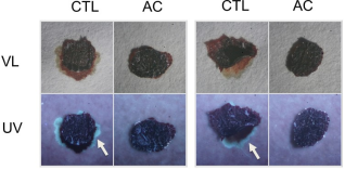
Kearse’s controlled experiments found that 365-nanometre ultraviolet illumination can reveal serum-associated patterns and that blood allowed to clot on skin before transfer to absorbent material can produce fluorescent peripheral borders. Anticoagulated samples that did not form a normal clot lacked comparable borders in the illustrated setup. This supports the mechanism that prior coagulation and fluid separation can produce an ultraviolet-visible margin. It does not establish that every Shroud margin is serum, nor that natural clot transfer is the only way to create a similar distribution.18, 20
Kearse also studied how temperature and humidity affect serum patterns while fresh blood dries. In high humidity, normal coagulation can be disrupted and the characteristic peripheral pattern may be reduced or absent. This is distinct from his separate experiment in which already-dried stains were exposed to high humidity and later transferred. The two mechanisms must not be conflated: humidity can affect initial clot formation and can, under other conditions, rehydrate a dried stain.21, 22
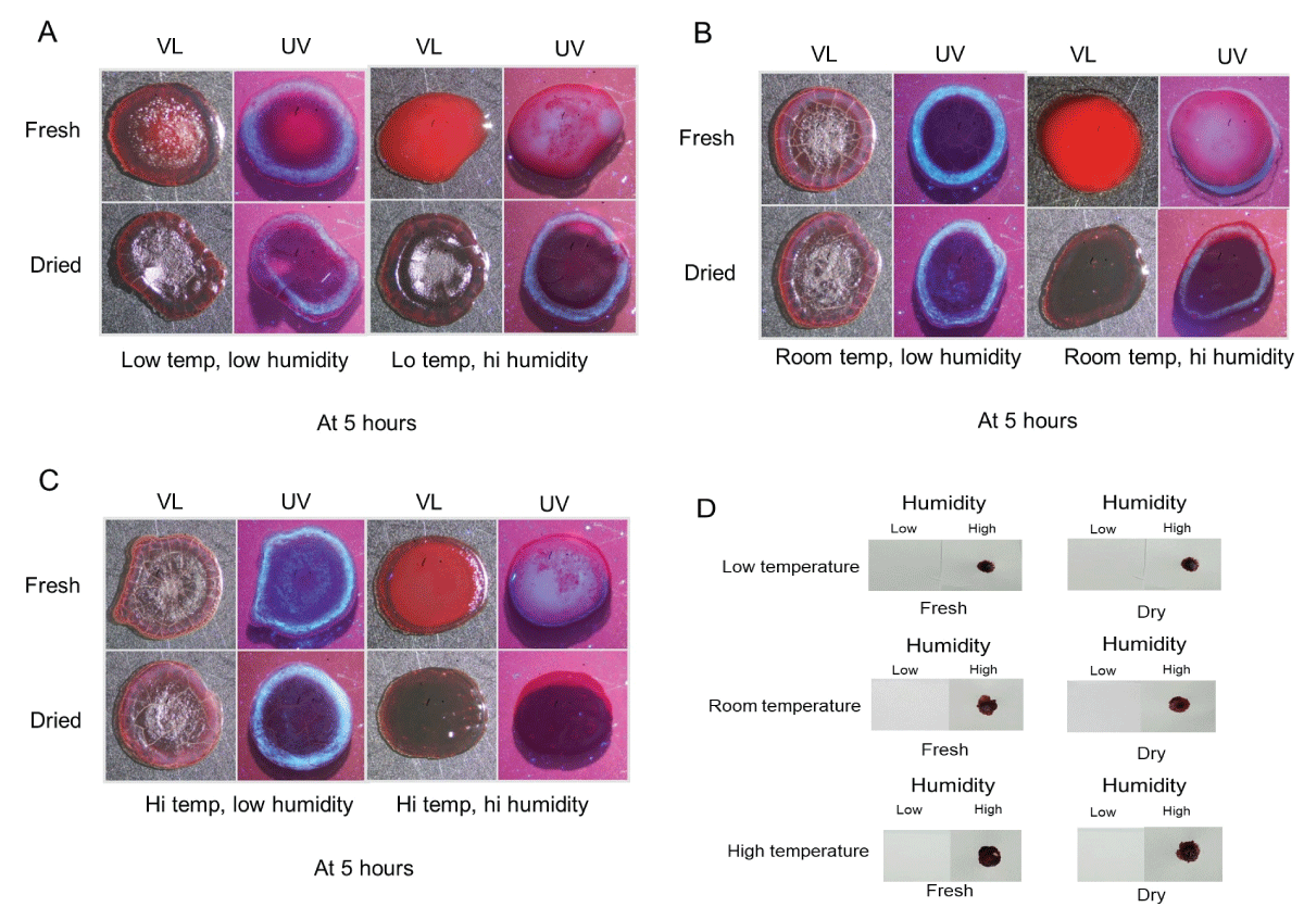
Even if a margin is protein-rich or serum-associated, several explanations remain possible: clot retraction before transfer, plasma or serum added separately, a different biological fluid, water-mediated migration, or deliberate application. A halo can support prior separation or differential wicking, but its unique cause has not been validated on the ancient stains. It cannot serve as a clock by itself.
8. How blood can transfer to linen
One long-standing objection to a body-to-cloth transfer is that a dried clot might not leave a clear image. Gilbert Lavoie and colleagues explored the timing of clot transfer by placing linen onto blood clots at different intervals. In their laboratory setup, recognizable transfer diminished as the clot dried; the time window varied with conditions. These experiments establish that transfer is sensitive to timing, moisture, pressure, and material. They do not recreate a burial or show that the Shroud stains formed in the same way.23
Kearse’s humidity experiments address a different possibility. Dried blood on skin or another non-absorbent surface can, under sufficiently humid conditions, regain a gelatinous state and transfer to absorbent material with recognizable edges. That is a demonstrated laboratory possibility under the tested conditions. It does not prove that a particular tomb was humid enough, that a specific body remained in such an environment, or that this is how the Shroud marks were made. The author’s later review correctly removes the often-repeated claim that the relevant tomb can simply be assigned a precise temperature of about 10 °C and relative humidity of 95 percent; no measurement of that specific ancient tomb establishes those values.22, 24
A natural transfer model must account for several physical variables: whether the blood was liquid, clotted, or partly dried; how much pressure brought cloth into contact with skin or wound; whether hair, folds, or body contours interrupted contact; the direction of gravity; how the body was moved; the weave and absorbency of the linen; and the environmental conditions. Experimental results demonstrate possible mechanisms, not a unique historical event.
9. Postmortem flow, washing, and humidity
Death stops active circulation, but it does not instantly make all blood solid. Blood may clot, remain fluid, or later become less coagulable through postmortem fibrinolysis. Raymond Mole’s classical paper and later work describe the relation between fibrinolysis and postmortem blood fluidity. Frederick Zugibe discussed passive oozing from disturbed or washed wounds. A fluid-looking stain can therefore be compatible with postmortem drainage, but fluidity is not diagnostic of one cause of death. It does not prove asphyxia, nor does it show that the heart continued to pump.25, 26, 27
A postmortem flow, if present, would be passive: gravity, body rotation, tissue compression, opening of a wound tract, release of pooled blood, or mechanical disturbance could move fluid after death. Unnailing and handling are plausible contexts to test, but no complete experiment has reproduced the Shroud wrist sequence from a realistic nail wound through death, removal, movement, contact, and linen transfer.
The author’s earlier *Forensic Summary* states a stronger contrast: some marks show serum halos and are therefore premortem, while other flows are postmortem. The later 2026 review appropriately qualifies this. No validated Shroud-specific assay has conclusively sorted ancient marks into premortem and postmortem categories. Some stains may be compatible with clot-associated transfer, and some rivulets may be compatible with later passive flow, but assigning a timeline from morphology alone is an inference with substantial uncertainty.27, 28
Claims that the body was or was not washed face the same problem. Washing could remove or redistribute some materials; burial practices and the condition of a violently injured body are historically complex. Stains that remain visible do not prove that no washing occurred, and a missing stain does not prove washing. Kearse’s 2025 paper revisits washing as a transfer hypothesis, but a definitive answer would require controlled experiments on realistic wounds and textiles, plus a reliable map of the relevant ancient marks.29
10. What bloodstain-pattern experiments can tell us
Bloodstain-pattern analysis examines direction, shape, distribution, and transfer. In 2019, Matteo Borrini and Luigi Garlaschelli tested selected Shroud interpretations using volunteers, simulated blood, and a mannequin. They argued that some commonly proposed flows did not fit the particular postures and application methods they tested. Their work is a challenge to overly confident reconstructions, not a complete simulation of crucifixion, death, unnailing, transport, and burial.30
In 2024, Andreas Heger and colleagues investigated wrist and forearm flow patterns while living volunteers changed arm positions. Their results show that movement can redirect flows and generate branches in some setups. The experiment did not model an actual nail tract, a cadaver, postmortem fibrinolysis, or transfer to linen. A separate 2024 study by Lutz König and co-authors examined proposed stains attributed to the crown of thorns, side wound, and a blood belt, using experimental approaches to test formation hypotheses. These studies make particular geometries testable; they do not certify one reconstruction as the historical answer.31, 32
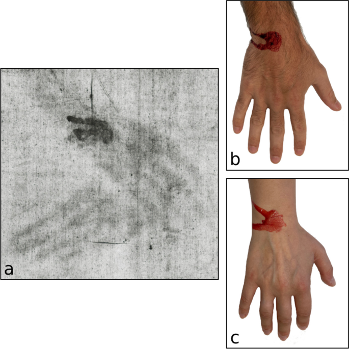
Experiments are strongest when their scope is explicit. They can show that a particular flow is possible or difficult under specified conditions. They cannot establish an event simply because a pattern resembles the experimental result. Realistic test programmes need transparent protocols, varied body positions, skin and hair, realistic wounds, measured blood properties, controlled humidity and temperature, multiple fabrics, blinded evaluation, and comparison against deliberate application using genuine blood as well as non-biological pigments.
11. A composite wrist-wound model—and its limits
The author’s 2026 critical review proposes that some wrist marks could combine more than one phase: bleeding while circulation continued; partial clot formation and serum separation; later drying; mechanical disturbance when a nail was removed or a body was moved; passive postmortem drainage; and contact with linen while some material remained moist or had been rehydrated. This is a coherent, testable model that can reconcile an older clot-associated component with a later liquid-looking flow. The individual mechanisms have experimental or forensic analogues, but the complete sequence has not been demonstrated in one anatomically realistic reconstruction.27
The model makes concrete predictions. A mapped wrist mark should contain spatially distinct components consistent with an older cellular-rich deposit and a later flow; the ultraviolet margin should not simply follow every part of the later rivulet; and the proposed sequence should outperform alternatives such as immediate wet contact, washing-mediated transfer, separate application of clear fluid, and artistic application of genuine blood. The test should be preregistered, blinded, and include negative controls. Until such work is done, “composite transfer” is a plausible explanatory hypothesis, not a forensic finding.
The same caution applies to the side wound and to claims that blood separated into red material and clear fluid in a way that uniquely confirms a Gospel account. Blood can separate after settling; serum or other fluids can move through cloth; and a stain image cannot independently establish the exact wound, its timing, or a scriptural event. The physical comparison is worth studying, but historical attribution requires evidence beyond fluid mechanics.33
12. What the evidence can support
Well documented: the cloth bears reddish marks in locations interpreted as injuries; STURP investigators recorded them photographically and sampled selected areas; Heller and Adler published chemical and spectroscopic findings interpreted as blood-related; McCrone published a pigment interpretation; and Miller and Pellicori documented ultraviolet fluorescence around some marks. Modern experiments show that clotting, serum separation, humidity, contact time, and movement can alter how blood-like materials transfer to absorbent fabric.2, 5, 8, 18, 22, 30, 31
Supported, but not uniquely established: some sampled stains may contain blood components; some fluorescent margins are compatible with serum-rich material; and mixtures of clot-associated transfer and later passive flow are physically plausible. The degree to which these findings characterize every visible stain remains uncertain.
Not established by the blood evidence: the donor’s species, sex, ABO group, genetic identity, exact cause of death, or identity as Jesus; the exact timing of each stain; a unique corpse-to-cloth transfer process; or the date and authenticity of the Shroud. These questions require independent, validated evidence. The blood evidence is important because it constrains possible explanations, not because it settles every historical question.
13. What would make the evidence stronger?
First, digitize the original 1978 visible and ultraviolet photographs with calibrated color and scale, then publish the unprocessed files and acquisition details. Second, map every candidate mark across the whole cloth, distinguishing stain core, pale margin, overlaps, repairs, and fire or water alteration. Independent observers should classify marks blind to the favored interpretation, and the study should report both positive and negative examples.
Third, build a controlled reference set using fresh and aged blood, serum, plasma, other biological fluids, pigments, and deliberately applied mixtures on linen of known weave. Vary clotting time, humidity, temperature, pressure, contact time, skin and hair, movement, washing, fire exposure, and later rehydration. Use modern assays with controls and independent laboratories. For the wrist hypothesis, model a realistic wound and postmortem handling sequence; compare it with immediate transfer and artistic application using genuine blood. Publish all images, protocols, failed trials, and error rates.
Finally, avoid using experimental compatibility as a proxy for identity. Even if a future study securely demonstrates blood-derived material and a particular sequence, it would not date the linen or identify whose blood it was. Those are separate evidence questions. The strongest next research is research that could prove the favored model wrong as well as support it.
References and research papers
- Giorgio Frache, Eugenia Mari Rizzati, and Emilio Mari, “Relazione conclusiva sulle analisi di ordine ematologico effettuate sul materiale prelevato dalla Sindone” (1976), Turin commission report. The tests were reported negative on the material examined; the result does not establish that all Shroud stains are blood-free. [Report transcription and context].
- John H. Heller and Alan D. Adler, “Blood on the Shroud of Turin,” Applied Optics 19, no. 16 (1980), pp. 2742–2744. [DOI] · [Paper copy].
- John H. Heller and Alan D. Adler, “A Chemical Investigation of the Shroud of Turin,” Canadian Society of Forensic Science Journal 14, no. 3 (1981), pp. 81–103. [DOI] · [Paper copy].
- E. J. Jumper, A. D. Adler, J. P. Jackson, S. F. Pellicori, J. H. Heller, and J. R. Druzik, “A Comprehensive Examination of the Various Stains and Images on the Shroud of Turin,” in Archaeological Chemistry III, ACS Advances in Chemistry 205 (1984), pp. 447–476. [Read the paper].
- Vernon D. Miller and Samuel F. Pellicori, “Ultraviolet Fluorescence Photography of the Shroud of Turin,” Journal of Biological Photography 49, no. 3 (1981), pp. 71–85. [Paper and photographs].
- S. F. Pellicori, “Spectral Properties of the Shroud of Turin,” Applied Optics 19, no. 12 (1980), pp. 1913–1920. [DOI].
- Walter C. McCrone and Christine Skirius, “Light Microscopical Study of the Turin ‘Shroud’ I,” The Microscope 28 (1980), pp. 105–113. [Publication record].
- Walter C. McCrone, “The Shroud of Turin: Blood or Artist’s Pigment?” Accounts of Chemical Research 23, no. 3 (1990), pp. 77–83. [DOI].
- Giulio Fanti and G. Zagotto, “Blood Reinforced by Pigments in the Reddish Stains of the Turin Shroud,” Journal of Cultural Heritage 25 (2017), pp. 113–120. [DOI].
- Gian Franco Di Minno, R. Scala, I. Ventre, and G. de Gaetano, “Blood Stains of the Turin Shroud 2015: Beyond Personal Hopes and Limitations of Techniques,” Internal and Emergency Medicine 11, no. 4 (2016), pp. 507–516. [DOI].
- A. Di Lascio, P. Di Lazzaro, P. Iacomussi, M. Missori, and D. Murra, “Investigating the Color of the Blood Stains on Archaeological Cloths: The Case of the Shroud of Turin,” Applied Optics 57, no. 23 (2018), pp. 6626–6631. [Publisher record].
- Kelly P. Kearse, “Unanticipated Issues in Serological Analysis of Blood Species: The Shroud of Turin as a Case Example,” Forensic Science International: Reports 2 (2020), article 100073. [Open-access paper].
- Kelly P. Kearse, “Major Drawbacks of Blood Species Analysis Using Human Polyclonal MNS Antisera: The Turin Shroud as a Case Example,” International Journal of Forensic Sciences 9, no. 4 (2024). [DOI].
- Pierluigi Baima Bollone, M. Jorio, and A. L. Massaro, “Identificazione del gruppo delle tracce di sangue umano sulla Sindone,” Sindon: Quaderno 31 (1982), pp. 5–9. Early report of ABO-group testing, discussed critically in reference 15.
- Kelly P. Kearse, “Inadequacies in Serological ABO Typing of Ancient Artifacts: The Shroud of Turin as a Case Example,” International Journal of Forensic Sciences 8, no. 3 (2023). [DOI and article].
- Kelly P. Kearse, “Validation and Limitations in the DNA Analysis of Aged Bloodstains: The Shroud of Turin as a Sample Case,” Forensic Science International: Reports 8 (2023), article 100335. [DOI].
- Louis A. Schwalbe and Raymond N. Rogers, “Physics and Chemistry of the Shroud of Turin: A Summary of the 1978 Investigation,” Analytica Chimica Acta 135 (1982), pp. 3–49. [DOI].
- Kelly P. Kearse, “Ultraviolet 365 as an Alternative Light Source for Detection of Blood Serum,” Journal of Forensic Sciences 65, no. 5 (2020), pp. 1716–1721. [DOI].
- Kelly P. Kearse, “Visualization of Plasma and Serum Pattern Formation in Drying Drops of Blood,” ARC Journal of Forensic Science 6, no. 1 (2022), pp. 1–5. [Article].
- Kelly P. Kearse, “Ultraviolet Fluorescent Detection of Elevated Bilirubin in Dried Blood Serum,” Journal of Forensic Science and Research 6 (2022), pp. 49–52. [DOI].
- Kelly P. Kearse, “Environmental Influence on Blood Serum Detection Using Ultraviolet 365,” Journal of Forensic Science and Research 5 (2021), pp. 30–36. [DOI].
- Kelly P. Kearse, “The Effect of Humidity on Blood Serum Pattern Formation and Blood Transfer,” Journal of Forensic Science and Research 7 (2023), pp. 40–48. [DOI].
- Gilbert R. Lavoie, B. B. Lavoie, V. J. Donovan, and J. S. Ballas, “Blood on the Shroud of Turin: Part II—The Importance of Time in the Transfer of Blood Clots to Cloth as Distinctive Clot Images,” Shroud Spectrum International 8 (1983), pp. 2–10. [Read the paper].
- Kelly P. Kearse, “A Simple, Natural Mechanism for the Transfer of Dry Bloodstains onto the Shroud of Turin,” International Journal of Archaeology 11, no. 2 (2023), pp. 17–21. [DOI].
- R. H. Mole, “Fibrinolysis and the Fluidity of the Blood Post Mortem,” Journal of Pathology and Bacteriology 60, no. 3 (1948), pp. 413–427.
- K. Haba et al., “Post-Mortem Activation of Human Blood Fibrinolytic Enzyme in Sudden and Natural Deaths,” Acta Medicinae Okayama 17 (1963), pp. 279–288.
- Otangelo Grasso, “Blood Transfer, Serum Halos, and Postmortem Flow on the Shroud of Turin: A Critical Forensic and Hematological Narrative Review” (2026). The author’s revised composite model, confidence grading, and experimental proposal. [Download the review].
- Frederick T. Zugibe, The Crucifixion of Jesus: A Forensic Inquiry (M. Evans, 2005). Forensic discussion of crucifixion injuries and postmortem flow.
- Kelly P. Kearse, “Blood Transfer to the Shroud of Turin: The Washing Hypothesis Revisited,” International Journal of Archaeology 13, no. 2 (2025), pp. 152–156. [DOI].
- Matteo Borrini and Luigi Garlaschelli, “A BPA Approach to the Shroud of Turin,” Journal of Forensic Sciences 64, no. 1 (2019), pp. 137–143. [DOI] · [Author manuscript].
- Andreas Heger et al., “Further Experiments and Remarks Regarding the Possible Formation of Blood Stains on the Turin Shroud: Stains Attributed to the Nailing of the Hands,” International Journal of Legal Medicine 138 (2024), pp. 1573–1581. [DOI].
- Lutz König, R. Schmölders, M. Jähling et al., “Some Experiments and Remarks Regarding the Possible Formation of Blood Stains on the Turin Shroud: Stains Attributed to the Crown of Thorns, the Lance Wound and the Belt of Blood,” International Journal of Legal Medicine 138 (2024), pp. 229–238. [DOI].
- Otangelo Grasso, “Forensic Assessment of the Shroud of Turin and the Sudarium of Oviedo” (2026), supplied critical review of reported correspondences and their limits. [Download the article PDF].
- Otangelo Grasso, The Shroud of Turin: A Forensic Summary of the Evidence, 3rd ed., chapter 3, “The Blood of the Shroud”; and From Forensics to Faith (2026), blood and wound discussions. The book’s interpretations are distinguished here from the underlying observations and independently assessed claims. [Find Forensic Summary on Amazon] · [Find From Forensics to Faith on Amazon].
- Otangelo Grasso, “A Forensic and Biochemical Analysis of the Bloodstains on the Shroud of Turin: Synthesis of Macroscopic, Microscopic, and Stratigraphic Evidence” (2026). Author’s synthesis; some claims are qualified by the later review in reference 27. [Download the article PDF].
- Otangelo Grasso, “Pre-Mortem and Post-Mortem Blood on the Shroud of Turin: A Convergent Constraint Analysis” (2026). Author’s earlier synthesis, read alongside the later evidential qualifications in reference 27. [Download the article PDF].
The author’s supplied studies are also listed in the research collection and PDF article index.