IN-DEPTH READING
Crucifixion trauma: reading anatomy without overreading the image
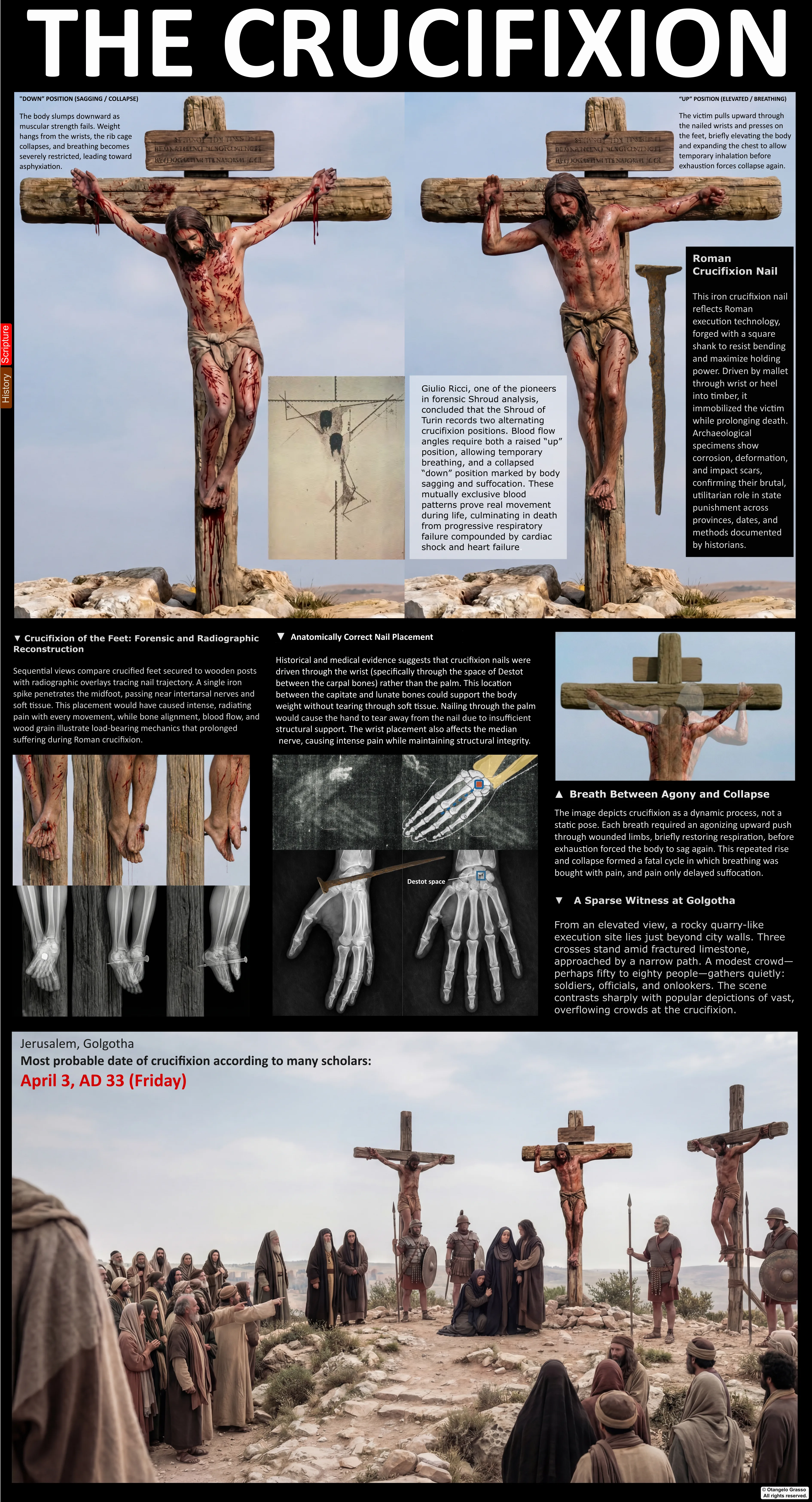
Crucifixion was a Roman form of public execution, but it did not have one fixed anatomical template. The position of the victim, the use of nails or bindings, the shape of the support, and the injuries inflicted before suspension could vary. This chapter examines the bodily injuries proposed for the man represented on the Shroud of Turin and asks what the textual record, archaeology, anatomy, experiments, and cloth image can each support.
Four evidence types must remain distinct throughout: text (what ancient authors report), archaeology (physical remains and excavated structures), observation (features visible or measured on the cloth), and interpretation (a proposed cause or mechanism). A mechanism that is anatomically possible does not prove that it happened. A mark interpreted as a wound is not automatically a chemically identified wound. A plausible reconstruction is a model, not an eyewitness record. The Shroud’s image formation and historical identity remain contested; this chapter does not assume either side of that debate.
1. What ancient texts say—and leave unsaid
The New Testament reports that Jesus was crucified, that soldiers divided his garments, that his legs were not broken, and that a soldier pierced his side after his death (Mark 15; Luke 23; John 19). John also reports scourging and mockery before the final handover. The accounts do not specify the precise geometry of the cross, whether the hands were nailed through palms or wrists, how the feet were fixed, the number or design of scourges, or the exact physiological cause of death.1
These silences matter. Later devotional images, medical reconstructions, and modern films can make a single version of the event feel familiar, but familiarity is not independent historical evidence. The Gospel narratives should be treated as ancient literary sources with their own emphases and differences; the Shroud’s stains should be evaluated separately rather than used to fill every textual gap.
2. Roman practice and the limits of the physical record
Ancient references to crucifixion describe a range of punishments and bodily arrangements. Survivors, condemned people, enslaved people, rebels, and others could be treated differently. Posts, crossbeams, ropes, nails, and combinations of restraint are all discussed in the literature. There is no surviving Roman execution manual that prescribes a universal placement for every nail or a single standard posture.
The best-known first-century archaeological case is the man from Giv‘at ha-Mivtar near Jerusalem. A nail remained through the right heel bone, showing that at least one crucified person was fixed through the calcaneus. The skeletal evidence is incomplete and was reappraised by Zias and Sekeles; it cannot establish a standard method for fastening hands, the shape of a cross, or the method used in Jesus’s case.2 This individual burial is valuable precisely when its reach is kept narrow.
Modern reviews by Maslen and Mitchell emphasize how little direct archaeological evidence is available and how much reconstructions depend on assumptions. Their discussion also cautions that experimental demonstrations, clinical analogies, and retrospective diagnoses cannot recreate the full conditions of an ancient execution.3 Archaeology gives a real example; it does not provide a universal diagram.
3. Scourging: injury pattern versus instrument and count
Roman scourging could cause severe injury, but the short Gospel notices do not describe the instrument, duration, number of blows, or the victim’s condition afterward. “Flagrum” is often used broadly in modern accounts for a Roman whip; reconstructions that add multiple weighted thongs, metal pieces, or bones are not descriptions supplied by the Gospel text. Different implements and techniques could produce different marks and levels of tissue damage.
The Shroud’s dorsal image has been interpreted as showing many small marks and paired or grouped traces across the back and limbs. Researchers disagree about which marks are scourge injuries, how many are distinguishable, and whether the image resolves individual impact events. Counts such as “one hundred” or “more than one hundred” depend on choices about what qualifies as a separate mark, whether marks overlap, and how the image is enhanced. Such counts should be reported as an interpreter’s estimate, not a direct measurement of blows.
Even when a mark appears compatible with blunt or tearing trauma, the image alone cannot identify a specific whip, the force applied, the number of executioners, or the amount of blood lost. A high-resolution image, ultraviolet photographs, and microscopic examination can clarify where traces appear, but chemical identification, chronology, and causal attribution require separate evidence. The owner’s papers on image requirements and consolidated interpretation are included among the author’s working analyses; they are not substitutes for independent experimental data.4
4. Head and scalp injuries
Some interpreters identify puncture-like marks around the head and propose an object made from thorn-bearing branches. The Gospels mention a crown of thorns in the mockery scenes, but do not describe its botanical species, shape, construction, or the number and distribution of punctures. A “cap” or helmet-like arrangement is one reconstruction; a circlet or another form remains possible. The faint head image, hair, blood-like traces, and image distortion make precise counting especially uncertain.
Small stains near the scalp have also been used to infer particular vessels, directions of flow, or repeated positions. Those conclusions require several steps: first deciding that a mark is a wound trace; then identifying the fluid and body location; then inferring posture and timing. Each step carries uncertainty. The stains do not, by themselves, identify a species of thorn or a precise sequence of movements. The author’s separate crown study evaluates circlet and helmet-like proposals using the stain distribution and head-cloth geometry; it is a reconstruction argument, not a botanical identification or direct observation of the lost object.17 Head trauma deserves attention as a proposed component of the image, but it cannot be converted into a detailed clinical chart.
5. Hands and wrists: several possible paths
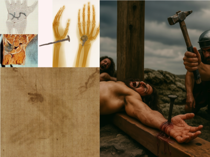
Pierre Barbet’s mid-twentieth-century work made the mechanics of nailing a central question in Shroud interpretation. He argued that a nail through the palm would not support a suspended body and proposed a route through the wrist, often associated with the so-called space of Destot. His work is historically influential, but the proposed route is not a demonstrated Roman rule. The term “hand” in ancient texts can also cover the wrist region, so the wording of a source cannot settle the anatomical issue by itself.
The wrist is a compact region of bones, joints, ligaments, tendons, vessels, and nerves. A nail’s direction, diameter, and precise position would affect what it encountered. “Destot’s space” is best treated as one proposed pathway through this complex anatomy, not as a uniquely established socket. Barbet’s demonstrations were informative but had limitations: they did not reproduce all possible support arrangements, durations, bodily conditions, or execution practices. A test that shows tearing under one arrangement does not establish that every palm placement was impossible.
Bordes and colleagues examined proposed hand, wrist, and foot pathways using five adult male cadavers. Their anatomical work demonstrates that more than one pathway is possible and that the consequences depend on the exact route. It does not recreate prolonged live suspension, scourging, movement, or Roman practice, and it cannot identify a particular nail tract from the Shroud image.6
Accordingly, claims that a nail “must” have passed through one named space exceed what the evidence can show. The Shroud itself offers a faint image of hands and forearms; it is not a three-dimensional wound specimen. The interpretation of a wrist-region stain as a nail exit or entry point is reasonable to investigate, but remains distinct from directly observing a nail tract in tissue or bone.
6. The missing-thumb proposal and nerve injury
A familiar argument says that the thumbs are not visible because a nail damaged the median nerve, causing the thumbs to flex into the palms. This combines an uncertain visual observation with a specific neurological mechanism. The image is faint, the hands overlap, and the boundary between a hidden thumb and an indistinct one is difficult to establish.
Clinical anatomy complicates the classic account. Regan’s review argues that a median nerve lesion at the wrist would not straightforwardly produce the often depicted posture; the level and completeness of injury matter, and paralysis should not be treated as a predictable spasm.7 Other mechanisms, including proximal nerve effects or arm position, have been proposed. Neither the absent-thumb observation nor a specific nerve injury can be diagnosed from the cloth with confidence.
7. Feet: what the heel-bone find can and cannot tell us
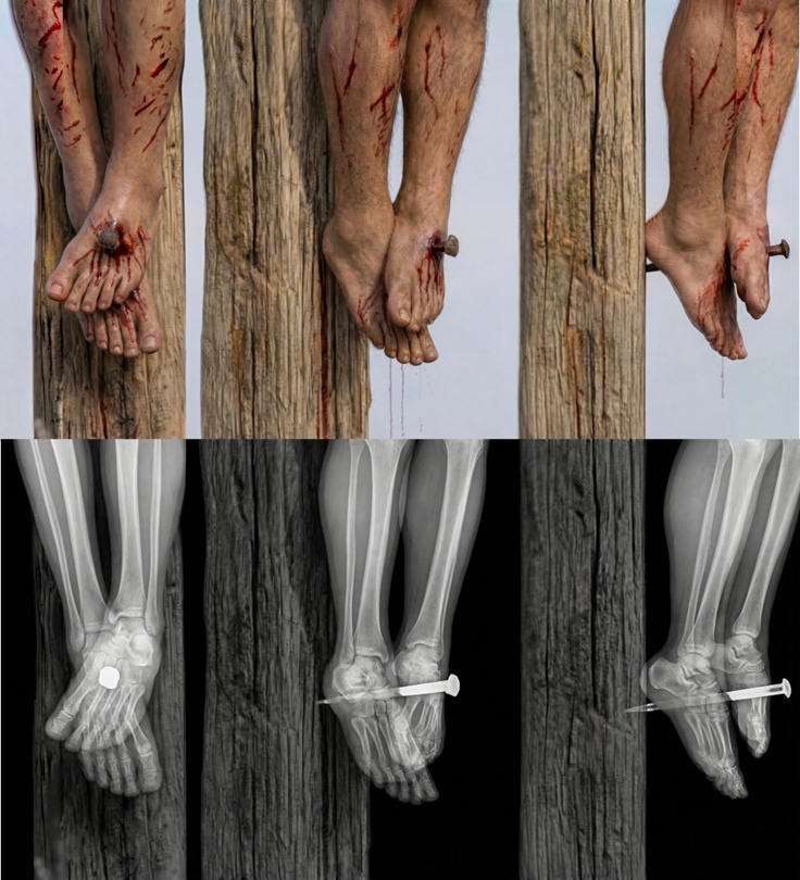
The Giv‘at ha-Mivtar burial preserves a nail through one heel bone. It is direct evidence that a nail could be used at the heel in at least one case. It does not tell us whether other executions used separate nails for each foot, one nail through overlapped feet, nails through the tarsal region, ropes, or another arrangement. The archaeological case is not a template for the Shroud figure.
Some Shroud interpretations propose that the feet were crossed and fixed by one nail; others discuss different positioning and pathways. Bevilacqua and colleagues combine image interpretation with anatomical testing to argue for a particular reconstruction. Their article is relevant as a Shroud-specific proposal, but it is not independent confirmation of the image’s age or identity, and its reconstruction remains one among debated models.8
The position of a body after removal from a cross may also differ from its position during suspension. Rigor, handling, limb movement, and the drape of cloth could alter the final geometry. The Shroud cannot establish the number of nails or the exact configuration from the apparent overlap alone.
8. Blood-flow directions and posture: the problem of non-unique explanations
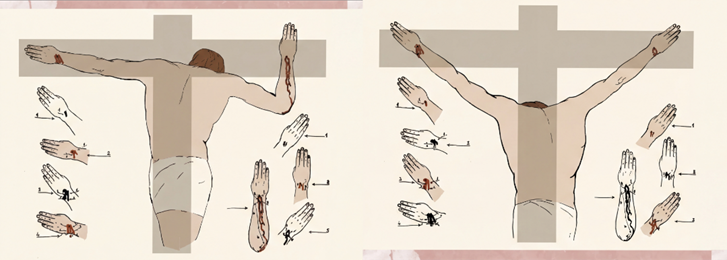
One influential interpretation reads different angles of wrist and forearm blood-like traces as evidence that the arms occupied changing positions. In the classic model associated with Barbet and later Shroud researchers, the victim periodically raised the torso, changing the shoulder and arm angle. This is a proposed physiological narrative, not a movement directly recorded by the cloth.
Other interpreters have challenged that account. Zugibe argued that a fixed arm position could produce the observed flow and proposed that an additional line might reflect events after death, such as removal or handling. Borrini and Garlaschelli tested several positions and concluded that not all marks fit a single posture in their experimental setup. Those experiments are useful countertests, though they too have limits: they do not reproduce actual penetrating wounds, all forms of restraint, prolonged suspension, or the complete history of the cloth.9
More recent experiments by König and colleagues examined the formation of wrist-region patterns under different arm positions and movements. Their work shows that similar flow directions can be generated under more than one arrangement, while noting the constraints of experimental models. A pattern that can be reproduced is not thereby proven to have formed in the same way historically.10 The question is underdetermined: gravity, arm rotation, wound location, contact with skin or fabric, fluid properties, movement, and later handling can all influence a stain.
9. Blood stains, serum halos, and identification claims
Shroud researchers have long disagreed about the chemistry and origin of the reddish-brown marks. STURP investigators reported findings they interpreted as blood-related, while McCrone argued that the material was pigment. Later proponents have discussed hemoglobin derivatives, proteins, and serum-like halos. These claims should be cited to the relevant analytical methods and samples rather than summarized as an uncontested consensus.
The location and appearance of a stain do not identify the fluid by themselves. A pale margin around a dark mark may be compatible with separation of components in a fluid, but appearance alone cannot establish serum chemistry, living circulation, or postmortem timing. The author’s biochemical review assembles macroscopic, microscopic, and stratigraphic arguments for a blood interpretation, but these are the author’s synthesis of debated analyses and are not a new independent laboratory result.18 Kearse’s experiments examine UV detection of serum-associated patterns, humidity, and blood transfer; Heger and colleagues later tested wrist-flow formation under different positions. These studies show that transfer and flow patterns depend on conditions and do not uniquely identify one event. A halo is therefore a feature to investigate, not a unique signature of one event.11
Blood type claims, including the frequently repeated “AB” classification, have also been debated because the available material is old, limited, and affected by contamination and sampling constraints. Even a confident identification of human blood would not by itself identify a person, date the stain, or establish the cloth’s provenance. The article “Blood Transfer, Serum Halos, and Postmortem Flows” by the author surveys this debate; it is a synthesis and should be read alongside the underlying laboratory literature, not as a new independent test.12
10. Lower-abdominal contour and suspension physiology
The frontal image has been described as showing a lower-abdominal prominence. Barbet connected the contour with respiratory effort during suspension; Zugibe disputed the classic asphyxia model while also discussing abdominal shape. In the author’s review, altered thoracoabdominal mechanics are proposed as one possible explanation, while cloth drape, image formation, body position, and postmortem change remain alternatives. The contour is not a diagnosis and no published crucifixion experiment directly measures it on a historical body.13
A useful distinction is between compatibility and specificity. A feature may be compatible with a proposed posture without being specific to that posture. Since the same apparent contour may result from several factors, it cannot independently establish suspension, cause of death, or authenticity.
11. Side wound and the boundary with the next chapter
John 19:34 reports that a soldier pierced Jesus’s side after death and that blood and water appeared. Some researchers interpret a side-region mark on the Shroud as a lance wound with a flow pattern. This chapter records the anatomical issue but does not treat the Gospel phrase as a clinical description. The image cannot identify the weapon, exact entry point, injured organs, or the physiological source of fluid.
Proposed explanations include injury to the chest wall, pleural space, pericardium, or heart, but these are retrospective scenarios without a body or sample to examine. Chapter 13 addresses the side wound and its medical interpretations in detail; keeping that analysis there avoids turning a visual resemblance into a diagnosis here.1
12. Cause of death: no single mechanism established
Published proposals include respiratory impairment, shock from trauma and blood loss, exhaustion, cardiac disturbance, and combinations of mechanisms. The popular image of a simple “asphyxiation cycle,” in which the victim must repeatedly push upward to breathe, is one model rather than a settled medical finding. The position, duration, supports, injuries, hydration, temperature, and individual condition would matter, and those details are unknown for the person represented on the Shroud.
Maslen and Mitchell’s review judged the evidence insufficient to establish one exact cause of death for crucifixion generally. Later reviews have argued different emphases, including whether suffocation was primary or whether other mechanisms better fit the available record. These are retrospective medical debates, not observations from a case file. The Gospels narrate that Jesus died; they do not supply the information required for a modern forensic cause-of-death determination.3
13. Evidence-weighted summary
| Evidence class | Supported conclusion | Not established by it alone |
|---|---|---|
| Gospel narratives | The texts report crucifixion, scourging, mockery, and a postmortem side-piercing episode in John. | Exact nail paths, wound dimensions, instrument design, or modern medical diagnosis. |
| Giv‘at ha-Mivtar | At least one first-century crucified man was nailed through a heel. | A universal Roman method or the specific procedure used in Jesus’s execution. |
| Cadaver anatomy | Several hand, wrist, and foot nail pathways are anatomically possible. | Which path was historically used, or how a living person would respond over time. |
| Shroud marks | Researchers identify features they interpret as wounds, fluid flows, and body contours. | Unique identification, exact movement, chemistry from visual appearance alone, or date. |
| Suspension experiments | Selected effects of constrained posture and arm position can be examined. | A complete execution, a single death mechanism, or the historical origin of a stain. |
| Author’s reconstructions | They organize hypotheses and make proposed mechanisms visible for discussion. | Independent confirmation unless supported by separate, reproducible evidence. |
The safest synthesis is modest. Roman crucifixion could inflict extensive trauma, and the Shroud bears marks that some researchers interpret as consistent with such an event. A heel nail in one archaeological burial and modern anatomical work make some proposed pathways physically possible. But the cloth is not a body, the historical method was not uniform, and multiple models can explain several of the same visual features. The exact instruments, nail routes, arm movements, timing of flows, and cause of death remain unresolved.
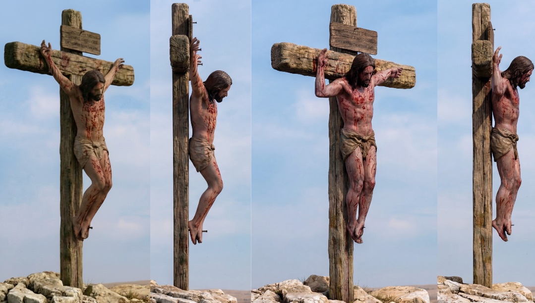
The images below and on the chapter panel are included to help readers inspect these proposals. They are supporting illustrations from the author’s book, not evidence independent of the hypotheses they depict. The author’s papers are identified as interpretive contributions; they should be weighed alongside the archaeological, anatomical, and medical sources listed below.
References and further reading
- The Gospel accounts: Mark 15; Luke 23; John 19:1–37. Selected passages.
- Zias, J., & Sekeles, E. (1985). “The Crucified Man from Giv‘at ha-Mivtar: A Reappraisal.” Israel Exploration Journal, 35(1), 22–27. See also “The Crucified Man from Giv‘at ha-Mivtar: A Reappraisal,” Biblical Archaeologist. Publication record.
- Maslen, M. W., & Mitchell, P. (2006). “Medical theories on the cause of death in crucifixion.” Journal of the Royal Society of Medicine, 99(4), 185–188. Full text.
- Grasso, O. (2026). A Consolidated, Illustrated Critical Assessment of the Shroud of Turin and Sudarium of Oviedo; and 12 Shroud of Turin Image Requirements and Assessment Criteria. Author’s analytical papers, identified as interpretive syntheses rather than independent experiments. Consolidated assessment; image requirements paper.
- Grasso, O. (2026). Forensic Summary, 2nd ed., ch. 12, pp. 214–226; Grasso, “Pierre Barbet and the Medical Study of the Shroud.” Author’s book chapter and review. Download the Barbet review.
- Bordes, S. J., et al. (2020). “The clinical anatomy of crucifixion.” Clinical Anatomy, 33, 12–21. Article.
- Regan, J. M., Shahlaie, K., & Watson, J. C. (2013). “Crucifixion and median neuropathy.” Brain and Behavior, 3(3), 243–248. Full text.
- Bevilacqua, M., et al. (2014). “How was the Turin Shroud Man crucified?” Injury, 45 Suppl. 4, S142–S148. Shroud-specific reconstruction. Article.
- Borrini, M., & Garlaschelli, L. (2019). “A BPA approach to the Shroud of Turin.” Journal of Forensic Sciences, 64, 137–143. Article record.
- König, M., et al. (2024). “Further experiments and remarks regarding the possible formation of blood stains on the Turin Shroud: stains attributed to the nailing of the hands.” International Journal of Legal Medicine, 138, 1573–1581. Article.
- Kearse, K. P. (2020). “Ultraviolet 365 as an alternative light source for detection of blood serum.” Journal of Forensic Sciences, 65(5), 1716–1721, doi:10.1111/1556-4029.14439; Kearse, K. P. (2023). “The effect of humidity on blood serum pattern formation and blood transfer.” Journal of Forensic Science and Research, 7, 040–048, doi:10.29328/journal.jfsr.1001048; Heger, A., et al. (2024). “Further experiments and remarks regarding the possible formation of blood stains on the Turin Shroud: stains attributed to the nailing of the hands.” International Journal of Legal Medicine, 138, 1573–1581, doi:10.1007/s00414-024-03166-7. These feasibility studies do not resolve provenance.
- Grasso, O. (2026). Blood Transfer, Serum Halos, and Postmortem Flow on the Shroud of Turin: A Critical Forensic and Hematological Narrative Review. Author’s synthesis; its proposed sequence is explicitly presented as plausible and experimentally testable, not uniquely proven. Read the review; related pre/postmortem analysis.
- Grasso, O. (2026). “Lower-Abdominal Protrusion in Crucifixion and the Turin Shroud: A Critical Review of Thoracoabdominal Mechanics, Suspension Physiology, Postmortem Change, and Three-Dimensional Image Interpretation.” Author’s review; written with AI assistance as disclosed in the source paper. Download the paper.
- Grasso, O. (2026). Forensic Summary, 2nd ed., ch. 12, pp. 214–226; illustrations reproduced from the author-supplied book file. They are explanatory reconstructions, not independent experimental evidence.
- Grasso, O. (2025). Crown of Thorns: Circlet or Helmet? A Forensic, Engineering, and Historical Analysis Integrating Cranial Wound Topography, Cloth–Head Geometry, and Relic Evidence. Author’s interpretive review, written with AI assistance. Download the paper.
- Grasso, O. (2026). A Forensic and Biochemical Analysis of the Bloodstains on the Shroud of Turin: Synthesis of Macroscopic, Microscopic, and Stratigraphic Evidence. Author’s synthesis, written with AI assistance; no new independent sample analysis. Download the paper.
- Grasso, O. (2026). Pre-Mortem and Post-Mortem Blood on the Shroud of Turin: A Convergent Constraint Analysis. Author’s interpretive analysis, disclosed as AI assisted. Download the paper.
- Grasso, O. (2026). STURP 1981 Symposium: Speakers’ Notes and Key Findings. Author’s synthesis of symposium proceedings, not the original proceedings. Download the notes.
- Heller, J. H., & Adler, A. D. (1980). “Blood on the Shroud of Turin.” Applied Optics, 19, 2742–2744. Article. Their analytical conclusion is historically important but remains part of a contested interpretation.
- McCrone, W. C. (1990). “The Shroud of Turin: blood or artist’s pigment?” Accounts of Chemical Research, 23(3), 77–83. Article.
- Lavoie, G. R., Lavoie, B. B., Donovan, V. J., & Ballas, J. S. (1983). “Blood on the Shroud of Turin: Part II—the importance of time in the transfer of blood clots to cloth as distinctive clot images.” Shroud Spectrum International, 8, 2–10. Clot-transfer experiments are analogues, not tests of the historical cloth.
- McGovern, M. E., Kaminskas, G., & Fernandes, J. R. (2023). “Did Jesus Die by Suffocation? An Appraisal of the Evidence.” Linacre Quarterly, 90(1), 64–79. Full text.
- Grasso, O. (2026). The Image That Should Not Exist: A Scientific Investigation of the Shroud of Turin, ch. 14, “The Anatomy of Crucifixion,” and ch. 15, “The Final Cascade.” Author’s synthesis; interpretation distinguished here from experimental and archaeological observations.